Welcome to MBC Biology
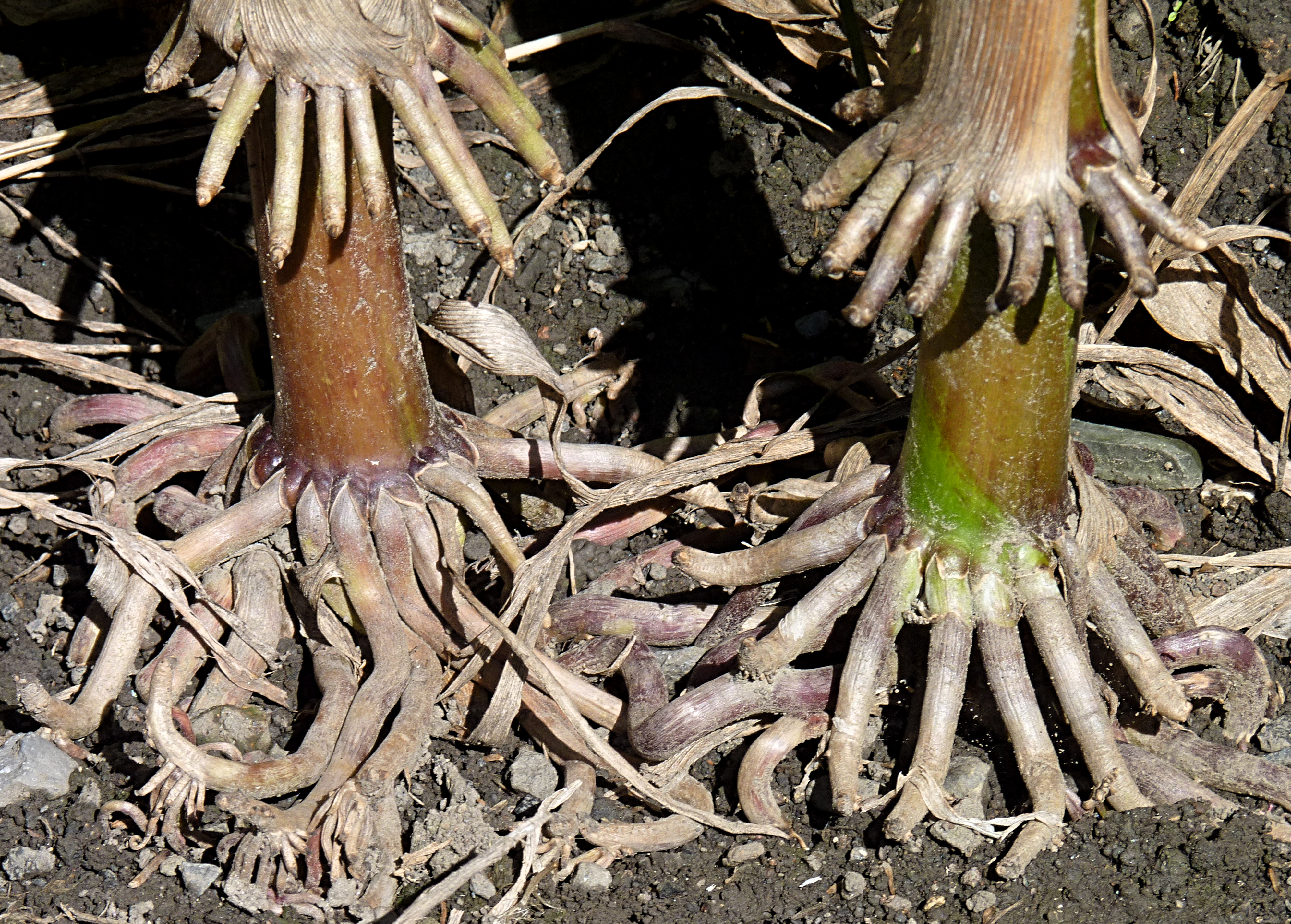 |
| Prop root of Maize (Zea mays) |
In monocot plants, a young and an old root do not have such distinction due to lack of secondary growth. A typical monocot root consists of epiblema, cortex, endodermis, pericycle, vascular bundles, conjunctive tissues, and pith (see Figure 13).
Epiblema
Epiblema is the outermost single layer of the root that is made up of compactly arranged, thin-walled, radially elongated parenchymatous cells without intercellular spaces. It does not have cuticle and stomata. Some cells elongate and make unicellular tubular outgrowths, called root hairs. As it has root hairs, epiblema is also called piliferous layer. Root hairs help in absorption of water and minerals from the soil.
Epiblema is the outermost single layer of the root that is made up of compactly arranged, thin-walled, radially elongated parenchymatous cells without intercellular spaces. It does not have cuticle and stomata. Some cells elongate and make unicellular tubular outgrowths, called root hairs. As it has root hairs, epiblema is also called piliferous layer. Root hairs help in absorption of water and minerals from the soil.
Cortex
Cortex lies below the epiblema. It consists of many layers of
thin-walled, oval, rounded or angular parenchyma with intercellular spaces. In
older roots, outer one layer (in Smilax) or many layers (in Zea mays)
of cortex contain sclerenchymatous tissues, called exodermis. Exodermis
gives mechanical support. Cortex helps in conduction of water from the root
hairs to the vascular system and storage of food.
Endodermis
Endodermis is the innermost
layer of cortex which is made of barrel-shaped compactly arranged
parenchymatous cells without intercellular spaces. Young endodermal cells lying
opposite to phloem possess an internal strips or bands of suberin and lignin,
called Casparian strip. A few thin-walled cells lying opposite to
protoxylem are called passage cells. Casparian strips do not allow the
movement of substances from cortex to pericycle. However, passage cells conduct
fluid inwards and outwards.
Pericycle
Pericycle lies below endodermis. It is made up of thin-walled
parenchymatous cells without any intercellular spaces. Pericycle may be
uniseriate (single layered in maize) or multiseriate (multilayered in Smilax).
It is responsible for the formation of lateral roots.
Vascular bundles
Vascular bundles are
made of xylem and phloem. Xylem and phloem are present in different bundles. They
are equal in number and lie alternate to each other. So, these vascular bundles
are called radial vascular bundles. Vascular bundles are more than six
(polyarchy) in monocot root.
Xylem is made of tracheids, vessels, xylem parenchyma, and
xylem fibres. Vessels are rounded or oval. Xylem bundles are exarch i.e. protoxylem
(the first formed xylem) lies towards periphery while metaxylem (the later
formed xylem) lies towards the centre of a root. Xylem helps in conduction of
water and minerals. It also provides mechanical support.
Phloem consists of sieve tubes, companion cells, rarely phloem
fibres. Phloem transports food.
Conjunctive tissue
One or many layers of
thin-walled parenchymatous or thick-walled sclerenchymatous cells are present in
between phloem and xylem, called conjunctive tissues. The parenchymatous
conjunctive tissues store food whereas sclerenchymatous conjunctive tissues
help in mechanical support.
Pith
Pith is the centre of
a root. In monocot root, pith is with large area. It is composed of
thin-walled, oval, rounded or angular parenchymatous cells with or without
intercellular spaces. It stores food.
 |
| Figure 13 a. T.s of monocot root (diagrammatic) |
 |
| Figure 13b. T. S. of monocot root (detailed view) |


No comments:
Post a Comment