Welcome to MBC Biology! In this post, I'm presenting you concise description of Epithelium.
Various
tissues combine to form an organ and different organs functioning together
constitute an organ system. Various systems constitute a body. Those tissues
are of four fundamental types (epithelial, connective, muscular,
and nervous tissue) based on their structure and function.
A.
Epithelial tissues
The
tissue which forms the outer and the inner coverings of various body parts
(skin), external parts, internal organs and body spaces inside are called epithelial
tissues or epithelium. The cells are closely packed; there is no
intercellular space or matrix exists between the cells. The cells are
held together by a cementing substance. Cells lie on a basement membrane,
which is not supplied with blood vessels. Epithelium contains nerve endings
too.
Based
on the shape of the cells and the layers, epithelium is simple, stratified
or compound, and modified epithelium.
1.
Simple epithelial tissues
Simple
epithelial tissue is composed of single layer cells resting on the basement
membrane. They are simple squamous, simple cuboidal,
simple columnar, and pseudo-stratified epithelium.
a.
Simple squamous epithelium
Simple
squamous epithelium is made of thin, flat, and hexagonal cells with a large
central rounded nucleus. The cells are closely packed like the tiles on the
mosaic floor. So, this tissue is also called pavement epithelium.
Simple
squamous epithelium is found in a covering around coelom, lining of the buccal
cavity, blood vessels, kidney, alveoli of lungs etc.
It
helps in protection, absorption, filtration, and exchange of gases.
b.
Simple cuboidal epithelium
Simple
cuboidal epithelium is composed of cuboidal cells having a centrally located
small rounded nucleus in each. Cells lie on a basement membrane.
It
is found in a lining on kidney tubules, sweat glands, salivary glands, gut,
testis, and ovary.
It
is involved in secretion, absorption, and excretion.
It
is of two types – ciliated cuboidal and brush bordered cuboidal epithelium.
Ciliated
cuboidal: Ciliated simple cuboidal epithelium contains cells with cilia on
the free surface. Each cell is associated with secretory goblet cells. They are
located on the ducts of the nephron. Cilia help in conducting the mucus and
other substances.
Brush
bordered cuboidal: Cells in this epithelium have microvilli at the free
ends. They are located at the proximal convoluted tubules of the nephron.
c.
Simple columnar epithelium
Simple
columnar epithelium consists of tall and narrow cells that are placed side by
side forming a layer like a column. Each cell has a large nucleus situated at
the basal end. These cells lie on a basement membrane.
It
is found in the lining of the goblet cells of the stomach, gastric glands,
intestinal glands, gall bladder, ureter, and uterine wall.
It
is of two types – simple ciliated columnar and brush bordered columnar
epithelium.
Ciliated
columnar: Cells in ciliated columnar are with numerous cilia on the free
surface. They are associated with secretory goblet cells. They are located in the oviducts, respiratory
passages (bronchioles), etc.
Brush
bordered columnar: This simple columnar epithelium has cells with
microvilli at the free ends of the cells. They are found in the intestinal
mucosa.
d.
Pseudo-stratified epithelium
Pseudo-stratified
epithelium consists of the columnar cells. As the cells do not reach free
surface and their nuclei appear to be at different levels, they provide a false
multilayered tissue. These cells rest up on the basement membrane.
It
is found in the lining of trachea, large bronchi, and urinary bladder.
It
protects the dust particles entering our respiratory tract.
2.
Stratified or compound epithelial tissue
Stratified
or compound epithelial tissue is made of several layers of the epithelial
cells. It is multi-layered as it contains an upper layer and a lower layer of
cells. Cells in a lower layer multiply and give rise to the cells of the upper
layers. The cells of a lower layer are called germinative cells.
Stratified
or compound epithelial tissue are stratified squamous epithelium, stratified
cuboidal epithelium, stratified columnar epithelium, and transitional
epithelium.
a.
Stratified squamous epithelium
The
tissue in which the upper layer of cells consists of large, flat and polygonal
or squamous cells, but the cells of germinative layer are either cuboidal
or columnar is stratified squamous epithelium. The first formed
cells are cuboidal shaped; they are pushed towards the upper surface outwards
and become flattened squamous. Stratified squamous epithelium is of two
types – keratinized stratified and non-keratinized squamous
epithelium.
keratinized
stratified epithelium: The uppermost layer consists of dead cells and is
hardened due to the deposition of keratin, a protein. The deposition of
keratin makes the cell layer water proof. It is located on hair, claws, and
nails.
Non-keratinized
squamous epithelium: The uppermost layer consists of living cells without
keratin. The layer is wet due to the absence of keratin. It is found on wet
surfaces like buccal cavity, pharynx, oesophagus, and vagina.
b.
Stratified cuboidal epithelium
It
is a stratified epithelium in which the outermost layer consists of cuboidal
cells. However, the lower layer has germinative cells either columnar or
squamous.
It
is found on the lining of the ducts of sweat glands, salivary glands,
pancreatic gland, and female urethra, etc.
c.
Stratified columnar epithelium
The
outermost layer of this tissue consists of tall columnar cells. However, the
germinative cells are cuboidal shaped.
It
is found on the lining of the ducts of mammary glands, lining of
vasa-differentia, trachea, and bronchi.
d.
Transitional epithelium
Transitional
epithelium is composed of three or four layers of cells. Cells in the uppermost
layer are dome-shaped; the middle layer cells are club-shaped; and the basal
layer cells are cuboidal or rounded cells. It has the capacity to stretch and
relax.
It is found in the lining of the urinary bladder, ureters, and uterus, etc.
3.
Modified epithelium ####
Some
epithelial tissues are modified for the specialized functions. These are ciliated
epithelium, sensory epithelium, Germinal epithelium,
and glandular epithelium.
a.
Ciliated epithelium
Ciliated
epithelium contains modified columnar or cuboidal cells. Those cells have cilia
at their free surfaces. It forms the lining of the neck of uriniferous tubules,
sperm ducts, trachea, and bronchi, etc.
b.
Sensory epithelium
Some
of the columnar cells are modified with sensory fibres at their free surfaces.
They form the lining of the tongue and the nasal cavity.
c.
Germinal epithelium
The
modified cuboidal cells found in the lining of testes and ovary make germinal
epithelium. They can divide and develop as gametes (spermatozoa and ova) by
meiosis. The germinal epithelium forms the lining of the gonads (seminiferous
tubule of testis and lining of ovary).
d.
Glandular epithelium
The
modified columnar or cuboidal cells specialized for manufacture and secretion
of certain chemical substances make glandular epithelium. The glandular
epithelia form glands.
 |
| Figure 9. Modified epithelium: 9 A. Ciliated epithelium; 9 B. Sensory epithelium |
Glands
The
glands are grouped on the basis of number of cells present; the kind of
secretion and the duct present; the shape and complexity; the mode of
secretion, and the nature of secretion.
Glands based on the number of cells present
There
are unicellular and multicellular glands based on the number of cells present
in them.
- Unicellular gland: A single cell scattered in the columnar cells is unicellular gland. Examples are goblet cells or mucus secreting cells.
- Multicellular gland: It is made of many cuboidal cells that form many tubular invaginations. Examples are sweat glands and gastric glands.
Glands based on the kind of secretion and the duct present
These
are exocrine and endocrine glands.
- Exocrine glands: The glands which pour their secretions through the ducts are called exocrine glands. They secrete enzymes. Glands can be unicellular or multicellular (simple or compound). Examples are salivary, tear, gastric, and intestinal glands.
- Endocrine glands: The glands that do not possess ducts but pour their secretions directly into the blood vessels are called endocrine glands. They are also called ductless glands. They secrete hormones. Examples are pituitary, thyroid, and adrenal glands, etc.
Glands based on the shape and complexity
Glands
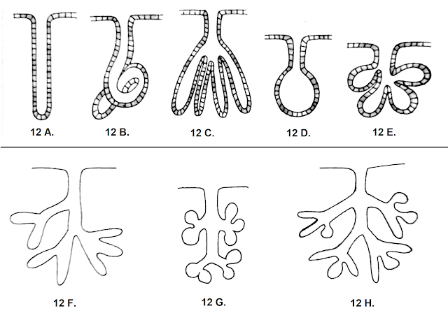
based on the shape and complexity or exocrine glands are of two types – simple
glands and compound glands.
Simple glands
These
glands have a single unbranched duct. The secretory part can be of tubular form
(called tubules) or sacs (called alveolar). These can be coiled
or uncoiled; branched or unbranched.
- Simple tubular glands are found in intestinal crypts in the intestine.
- Simple coiled tubular glands are found in simple sweat glands in the skin of mammals.
- Simple branched tubular glands are found in the linings of gastric glands and Brunner’s glands of intestine.
- Simple alveolar glands are found in the mucous secreting glands in the skin of frog.
- Simple branched alveolar glands are found in the sebaceous or oil glands in the skin of mammals.
Compound glands
These
glands have a number of ducts forming a branching pattern. The secretory part
can be in the form of tubes (tubules), sacs (alveoli), or both.
- Compound tubular glands are in the salivary glands.
- Compound alveolar glands are found in the mammary glands, pancreatic glands etc.
- Compound tubular-alveolar glands are found in the parts of salivary and mammary glands.
Glands based on the
mode of secretion
Depending
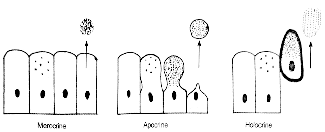
on the mode of secretion, the exocrine glands are of three types – merocrine
glands, apocrine glands, and holocrine glands.
- Merocrine glands: The secretions are discharged on the cell surface by diffusion or exocytosis without causing any damage or loss in the secretory cells. The cells remain intact e.g. goblet cells, salivary glands, intestinal glands, and sweat glands.
- Apocrine glands: The secretions are discharged on cell surface causing loss or damage to some parts of secretory cells e.g. mammary glands, eyelid, and ear, etc.
- Holocrine glands: The secretions are discharged on the cell surface by the rupture of the plasma membrane of secretory cells completely e.g. sebaceous glands in the skin of mammals.
Glands based on the nature of secretion
These
are mucous glands, serous glands, and mixed glands.
- Mucous glands: These glands secrete the mucus. The cells are called mucous cells or mucocytes. The mucus is a proteinous viscous and slimy substance. The goblet cells in the intestine are examples of mucous glands.
- Serous glands: These glands secrete a clear watery fluid. These cells are called serocytes. Serous cells are found in the parotid salivary glands, intestinal glands, and sweat glands.
- Mixed glands: Some glands are made of both the mucocytes and serocytes. These glands produce both kinds of secretions. Examples are gastric secretion and pancreatic secretion.
 |
| Figure 10 A. unicellular exocrine glands; 10 B. multicellular exocrine glands |
 |
| Figure 11 A. Exocrine gland; 11 B. Endocrine gland. |
 |
| Figure 13 Glands based on the mode of secretion |


No comments:
Post a Comment