Welcome to MBC Biology!
 |
| Image source: Needpix.com |
What is Tissue?
The term ‘tissue’ was coined by Nehemiah Grew, an English Anatomist and Physiologist, also known as ‘Father of Plant Anatomy’ in 1682. The term ‘tissue’ denotes a group of cells having a common origin and co-operating with one-another to perform a similar function.Plant tissues and their types
As the plants are multicellular organisms, they contain different types tissues. Based on the capacity of division, their tissues are of two types – meristematic tissues or meristem and permanent tissue.A. Meristems
- Meristems are composed of immature cells with capability to divide and grow.
- Intercellular spaces between cells are absent.
- Cells contain thin and elastic cell walls made of cellulose.
- Cells are oval, rounded, polygonal or rectangular.
- They do not store reserve food materials.
- The cells have dense cytoplasm with large, distinct and prominent nuclei.
- Vacuoles are either absent or very small.
- Metabolic activities are very high.
- Colourless proplastids are present.
Meristems are classified on the basis of origin and development,
position, and function.
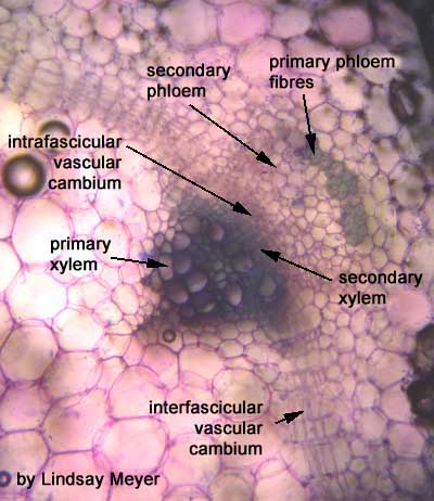
Secondary meristems are formed from the permanent tissues by dedifferentiation of permanent tissues. Examples are interfascicular cambium and cork cambium (See Figure1 c.).
II. Meristems on the basis of position
On
the basis of position, meristems are apical meristems, intercalary
meristems, and lateral meristems (See Figure 2).
1. Apical meristems
Apical
meristems are present at the tips or apex of stem, bud, root, and leaf. They
increase the length of plant.
2. Intercalary meristems
Intercalary
meristems are derived from apical meristems. They develop when permanent
tissues are formed in between apical meristems. They are found at the base of
leaves or at the bases of internodes.
3. Lateral meristems
Lateral
meristems are present along the sides of stem. Cells of these meristems divide
and increase thickness or girth of plant. An example is intrafascicular cambium
in dicot stem.
 |
Figure 2
L. S. of shoot showing the positions of meristems |
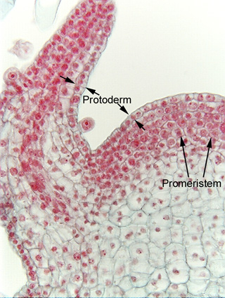
III. Meristems on the basis of function
Based on the function,
meristems are protoderm, procambium, and ground meristem
(See Figure 3).
1. Protoderm
Promeristem is the outermost layer of meristem. It gives
epidermis or epiblema of the developing parts of plants.
2. Procambium
Procambium lies internal to
the protoderm. It gives rise to primary vascular tissues.
3. Ground meristem
These are meristematic tissues except protoderm and procambium
in primary meristems. They make ground tissues such as hypodermis, cortex, endodermis,
pericycle, medullary rays, and pith.
B. Permanent Tissues
Permanent tissues are the tissues that have their growth has stopped either completely or they do not divide till their dedifferentiation happens. Sometimes, they regain meristematic activity partially or wholly. These tissues may be living or dead. The living permanent tissues have thin-walled or thick-walled cells whereas the dead permanent tissues have thick-walled cells. Permanent tissues are of two types – simple permanent tissues and complex permanent tissues.I. Simple Permanent Tissues
These
tissues are made up of one type of cells forming a uniform homogeneous system
of cells. Simple permanent tissues are of three types – parenchyma,
collenchyma, and sclerenchyma.
1. Parenchyma
Parenchyma is a tissue made of thin-walled living, similar,
oval, rounded or polygonal isodiametric cells with or without intercellular
spaces. Internally, cells have large central vacuole and peripheral cytoplasm,
and nucleus. It is found in the non-woody or soft areas of stems, leaves,
roots, flowers, and fruits etc. As they perform special function, they are
classified as epidermis, epiblema, simple parenchyma, chlorenchyma, aerenchyma,
prosenchyma, idioblast, phloem parenchyma, xylem parenchyma.
a. Chlorenchyma
Parenchyma having chloroplast are called chlorenchyma. They
are found in green stem, green fruits, and leaves. In leaves, they are called
mesophyll tissues. Mesophyll tissues are differentiated into palisade
parenchyma and spongy parenchyma (See Figure 4 a.).
b. Aerenchyma
Aerenchyma are parenchyma with air cavities. They are found in
aquatic plants and some land plants (xerophytes). The air cavities are
surrounded by thin-walled living oval, rounded, or irregular cells. Air
cavities store gases to make aquatic plants light and buoyant (See Figure 4 b.).
c. Prosenchyma
Prosenchyma are slightly thick-walled living fibre-like
elongated parenchyma (See Figure 4 c.). They are found in pericycle and
conjunctive tissues. They provide mechanical support and storage site of food.
Functions of Parenchyma
- Parenchyma helps in storage of food.
- They provide turgidity to softer parts of plants.
- Epidermis helps in protection of inner tissues.
- Chlorenchyma help in photosynthesis.
- Aerenchyma provide buoyancy and storage of metabolic gases.
- Phloem and xylem parenchyma helps in slow lateral conduction of materials.
- Prosenchyma help in mechanical support.
- Epiblema help in absorption of sap.
 |
| Figure 4 c. Prosenchyma tissue |
Collenchyma is a simple living permanent tissue with the cells having deposition of cellulose and pectin in specific areas of their wall. They are elongated, circular, oval or angular in transverse section. Internally, they possess large central vacuole and a peripheral cytoplasm along with nucleus. They may have a few chloroplasts. They are generally found in hypodermis of petiole, pedicel, leaf and stem of herbaceous dicot plants in the ridges; absent in woody dicot stem, monocot stem, and roots. Based on the thickening of cell wall, they are angular, lamellate, and lacunate collenchyma.
a. Angular collenchyma
They have wall thickening at
the angles (See Figure 5 a.). Examples are collenchyma in stem of tomato, Datura, Salanum,
Tagetes, etc.
b. Lamellate collenchyma
The wall thickenings are at
tangential walls (plate like thickening) (See Figure 5 b.). Examples are collenchyma in stem of
sunflower and Rhombus etc.
c. Lacunate collenchyma
The wall thickening is in the
intercellular spaces but with a small hollow cylinder (lacuna) (See Figure 5 c.). Examples are
collenchyma in stem of Cucurbita and petiole of Salvia etc.
Functions of Collenchyma
- Collenchyma provides mechanical strength to young stem, leaves, and petioles of herbaceous plant.
- It helps in cell elasticity and support to the growing organs.
- It provides support to delicate leaf margins and prevents tearing of leaves.
- It provides flexibility to organs and allows their bending. So, it prevents lodging of herbaceous dicot stem.
- It takes part in photosynthesis because it has chloroplast.
- It helps in storage of small amount of food.
- It allows growth and elongation of organs.
- Cells of collenchyma undergo dedifferentiation and can form cork cambium or phellogen.
3. Sclerenchyma
Sclerenchyma is a simple highly thick-walled dead tissue. The
wall is made up of cellulose or lignin or both. It is found in hard part of
plants. It provides mechanical support and stiffness to plants and their parts.
Sclerenchyma is of two types – fibres and sclereids.
a. Fibres
Fibres are highly elongated, narrow, and spindle-shaped thick-walled
dead cells with pointed end walls. They are arranged in longitudinal bundles. Fibres
occur in mechanical strength requiring parts like leaves, petioles, cortex,
pericycle, phloem, and xylem as well as around the vascular bundles (monocot
stem). Fibres are of three types – wood fibres (in xylem), bast fibres (in
phloem), and surface fibres (present in other than xylem and phloem) (See Figure
6 a.).
b. Sclereids
Sclereids are highly thickened dead sclerenchyma cells with
very narrow cavities. They are broader than fibres. They may be isodiametric,
polyhedral or cylindrical. The thick cell wall has branched or unbranched
simple pits. They occur singly or in groups. They provide stiffness to the
plants.
The different types of sclereids are stone cells or brachysclereids, macrosclereids, osteosclereids, astrosclereids and filiform sclereids (See Figure 6 b.). Brachysclereids give gritness to fruits of guava, apples, and pears etc. Macrosclereids are found in epidermal covering of some legume seeds. Osteosclereids are found in sub-epidermal covering of some legume seeds. Astrosclereids are found in tea leaves and petioles of lotus. Filiform sclereids are found in stem of hydrophytes.
Functions of Sclerenchyma
- Fibre type sclerenchyma help in mechanical support to the various parts of plants.
- Fibres allow the plant organs to tolerate bending, shearing, compression, and pull forces by environmental factors.
- Numerous fibres are commercially used e.g. Corchorus (Jute), Linus (Flax), Cannabis (Hemp), Agave, and Musa etc.
- Splitting and coiling of values during dehiscence of some fruits is due to orientation of sclerenchyma.
- Sclereids help in stiffness to the plant parts.
- Sclereids make stony endocarp of drupes, called stone fruits, in almond, and coconut etc.
 |
| Figure 6 a. Sclerenchyma tissue (fibre) |
 |
| Figure 6 b. Sclerenchyma tissue (sclereids) |
II. Complex Permanent Tissues
Complex permanent tissue is made up of different group of cells to perform a common function. So, it is heterogenous tissue. It is also called conducting tissue or vascular tissue, or physicomechanical tissue. It consists of phloem and xylem.
1. Phloem
Phloem
is a complex permanent tissue that transports food in plant (See Figure 7 a.).
It is also called bast. It consists of four types of cells – sieve elements,
companion cells/ albuminous cells, phloem parenchyma,
and phloem fibres.
a. Sieve elements
Sieve
elements are of two types – sieve tubes and sieve cells.
i. Sieve tubes
Sieve tubes
are elongated tubular conducting channels of phloem present in angiosperms (See
Figure 7 b.). They are placed end-to-end by transverse or oblique end
wall. They have small sieve pores. Each sieve pore is lined by a layer of
callose. Internally, a sieve tube has peripheral layer of cytoplasm without any
nucleus. The central part of sieve tube is occupied by a network of canals
which contain fibrils of protein. Sieve tubes take part in the conduction of
organic food.
ii. Sieve cells
Sieve cells
are the only conducting elements of phloem in pteridophytes and gymnosperms (See Figure 7
a.). Internally, a sieve tube or sieve cell has peripheral layer of
cytoplasm without any nucleus. Sieve cells take part in the conduction of
organic food.
b. Companion cells or
albuminous cells
Companion
cells are narrow, elongated and thin-walled living cells associated with sieve
tubes (See Figure 7 b.). They are present in the phloem of angiosperms. They
are square or rectangular in shape. Each companion cell has dense cytoplasm and
nucleus. In pteridophytes and gymnosperms, companion cells are replaced by albuminous
cells, the modified parenchyma. They are associated with sieve cells.
c. Phloem or bast parenchyma
Phloem
parenchyma is thin-walled living parenchyma without intercellular spaces and
associated with phloem (See Figure 7 b.). They help in storage and slow
lateral conduction of food.
d. Phloem fibres
Phloem fibres
are thick-walled dead sclerenchyma in phloem. They help in mechanical support
(See Figure 7 c.).
 |
| Figure 7 a. Phloem and sieve cell |
2. Xylem
Xylem is a complex permanent tissue that helps in transport of water and minerals. It also provides mechanical strength to plants. It has four components – tracheids, vessels, xylem or wood parenchyma, and xylem or wood fibres (See Figure 8).
a. Tracheids
Tracheids are
elongated thick-walled lignified dead cells with wide lumen and narrow end
walls. Their shape is polygonal or tetrahedral. These are only
conducting elements of xylem in gymnosperms and pteridophytes. In angiosperms, they are
present along with other conducting elements - vessels.
The walls of tracheids have annular (ring like), spiral (spiral or helix like), reticulate (network like), scalariform (ladder like), and pitted (uniformly thick except for small unthicken areas) thickenings for mechanical support. Annular is the most primitive while pitted is the most advanced thickening.
b. Vessels
Vessels are
much elongated tubes with either of the end closed. They are formed by union of
several short wide and thickened cells. Their end walls are transverse or oblique.
Their walls are lignified. They also have annular, spiral, reticulate,
scalariform, and pitted thickenings on the walls. Pitted thickening is more
common than others.
In T. S., they are circular in monocots whereas angular in dicots. They are absent in gymnosperms and pteridophytes.
c. Xylem parenchyma
It is thin-walled
living parenchyma without intercellular spaces. It helps in storage of water
and slow lateral conduction of sap.
d. Xylem fibres
Xylem fibres
are thick-walled dead fibre type of sclerenchyma. They help in mechanical
support.

Figure 8 a. T. S. of Xylem showing its elements

Figure 8 b. Thickenings in tracheids


 |
| Figure 8 c. Vessels with different end walls |
C. Special type of Tissues
Some tissues help in secretion or excretion, called secretory or excretory tissues. They are embedded in parenchyma of cortex, phloem, xylem or pith. Special types of tissues are laticifers or laticiferous tissue and Glandular tissues.I. Laticifers
- Storage of reserve foods like sugar, proteins, and oils;
- Formation and storage of excretory products like alkaloids, resins, tannins, and rubber etc.
- Conduction or translocation of products and regulate;
- Regulation of water balance in plants.
1. Latex cells
Latex cells
are branched, long individual cells which do not fuse to form a network (See Figure 9 a.). Examples
are latex cells in Calotropis, Euphorbia, Nerium, Vinca,
Cannabis, Urtica, Ficus, and Mulberry.
2. Latex vessels
Latex vessels
are long branched ducts which fuse to form a network (See Figure 9 b.). Examples are latex
vessels in Papaver, Argemone, Sunflower, Papaya, Banana, Cactus,
robber plant, etc.
 |
| Figure 9 a. Latex cell in plants |
 |
| Figure 9 b. Latex vessel in plants |
II. Glands or Glandular tissue
Glands
secrete oils, gums, mucilage, tannins, and resins. Glands are either external
or internal. External glands are hydathode (See Figure 10 a.), nectar glands,
glandular hairs, stinging hairs, digestive glands.
Internal glands are oil glands (See Figure 10 b.), mucilage glands,
and resin ducts, etc.
There may be three kinds of special cavities or canals – schizogenous, lysigenous, and schizo-lysigenous cavity - for the storage of their products (See Figure 11).
1. Schizogenous cavity
These
cavities are formed by separation of cells to leave an empty space. Examples
are resin ducts in stems of Pinus, Sunflower, Coriander, Fennels etc.
2. Lysigenous cavity
It is formed by break down of cells at particular spaces to
store aromatic oils or water. Examples are oil cavities of citrus, cloves, and Eucalyptus,
etc.
3. Schizo-lysigenous cavity
It is formed
by separation of cells and disintegration of cells. Example is protoxylem
cavity in maize stem.
 |
| Figure 11 Special cavities or canals surrounded by special tissues in plants |


No comments:
Post a Comment